Image credits
The radiographs and MRI on this site come from Wikimedia Commons under the licences below. They are shown cropped and graded in black and white; versions of images licensed CC BY-SA are shared under the same licence. Images inside the question bank belong to ST3 Interview.
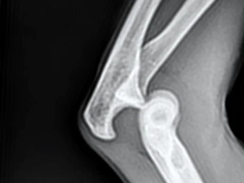
Posterior elbow dislocationRadiographie d’une luxation postérieur du coude gauche sans fractureFlibust1er · CC0
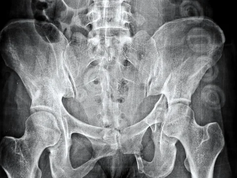
Pelvis, outlet viewPelvis outlet view showing fracture of the left ischiumCerevisae · CC BY-SA 4.0
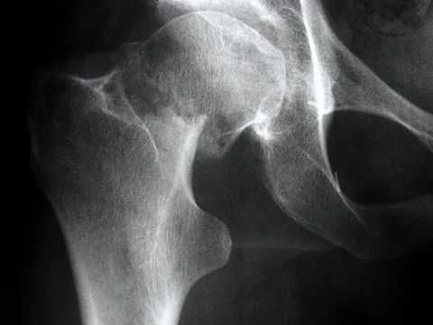
Fractured neck of femurFracture du col du fémurEucla, French Wikipedia · CC BY-SA 3.0
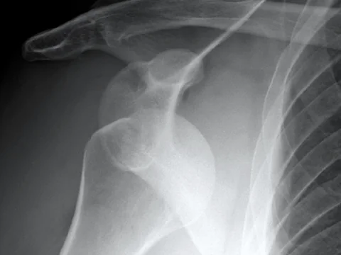
Anterior shoulder dislocation, APAnterDisAPJames Heilman, MD · CC BY-SA 4.0
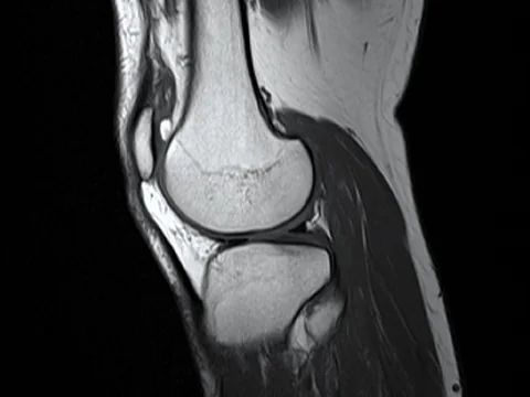
Knee MRI, sagittalKnee MRI T1 TSE SagittalPtrump16 · CC BY-SA 4.0
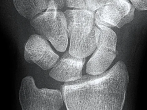
Scaphoid fractureScaphoidFrac2James Heilman, MD · CC BY-SA 4.0
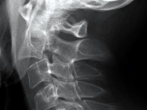
Hangman’s fracture, C2Hangman's fractureLucien Monfils · CC BY-SA 3.0
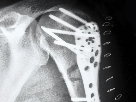
Plated proximal humerus fractureHumerus proximal winkstab 01aThomas Zimmermann (THWZ) · CC BY-SA 3.0 de
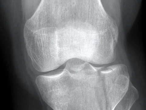
Lateral tibial plateau fractureLateral Tibial Plateau fracture XRay with Depression 2LottieLattes · CC BY-SA 4.0
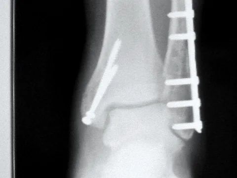
Ankle after fixationTrimalleolarFractureRepairR.SUNSET · Public domain
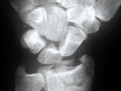
Distal radius fractureDisplaced distal radius fractureCurtishand · Public domain
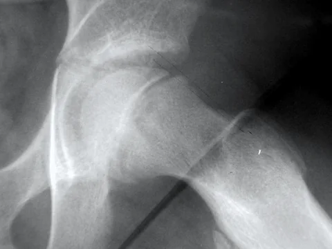
Slipped upper femoral epiphysisSCFE FROG B&WMikir at English Wikipedia · Public domain
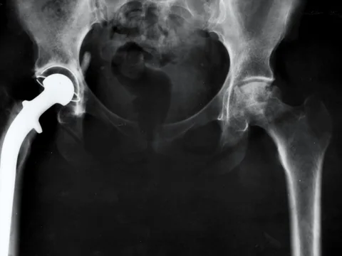
Hip replacementHip replacement Image 3684-PHUnknown photographer · Public domain
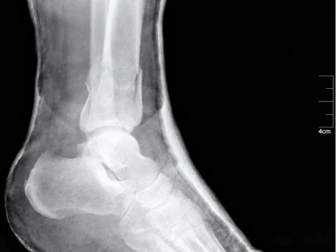
Ankle, lateralMedical X-Ray imaging NUW06 nevitNevit Dilmen · CC BY-SA 3.0
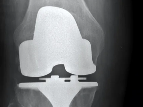
Total knee replacement, APRight Knee Post-surgery frontal implantUnknown · Public domain
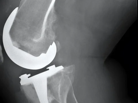
Total knee replacement, lateralRight Knee Post-surgery lateral implantUnknown · Public domain
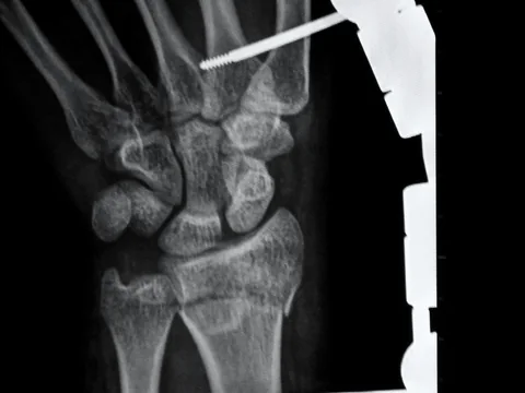
Wrist in an external fixatorExternal fixator xrayAshish j29 · CC BY 3.0

Portrait of Sam NahasSam Nahas · used with permission